Time: 18th-23rd June 2019
Place: ETH, Zurich, Switzerland
Participation fee: 500 CHF (academia) / 1'000 CHF (industry)

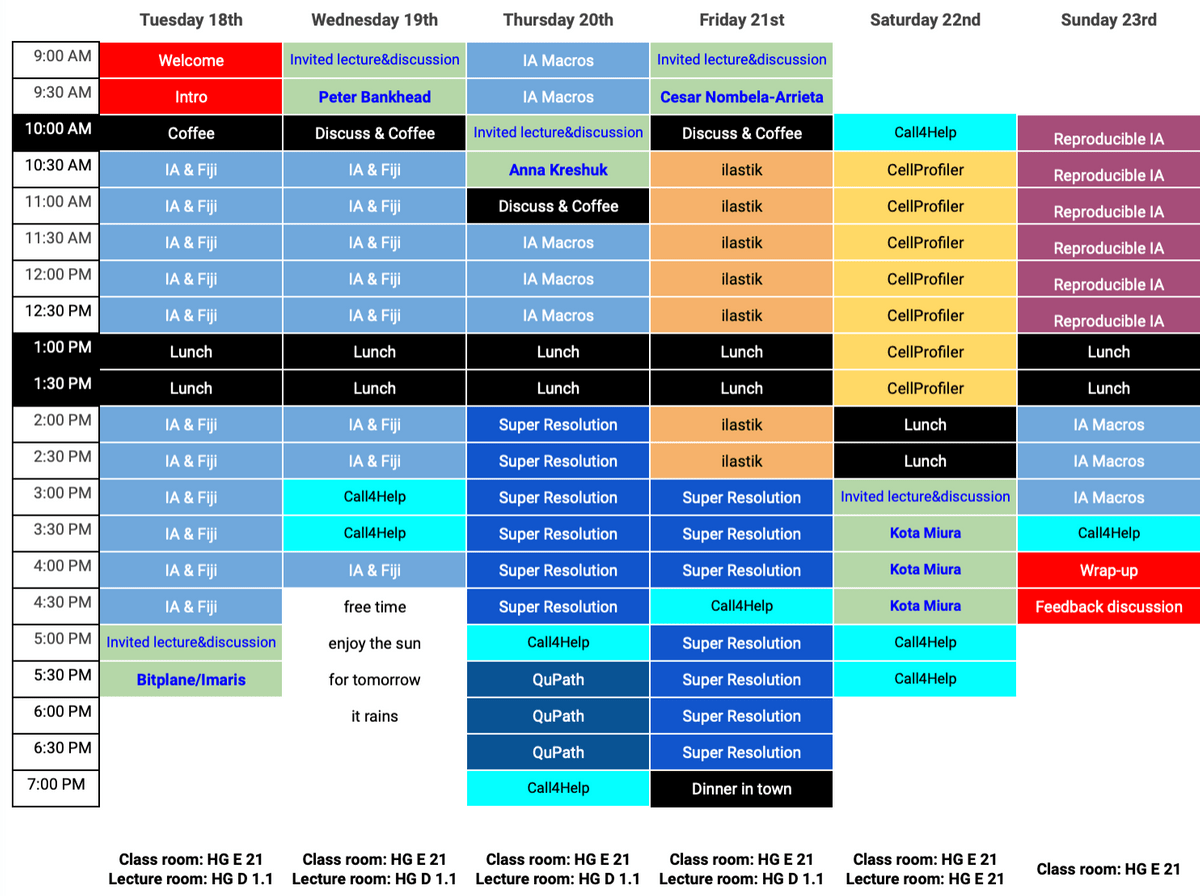
Program
Expect some changes as we adapt the pace dynamically

Full address: ETH Zürich; Main building; Rämistrasse 101; 8092 Zürich; Switzerland

Poster
Please help us promote the event!
Trainers
With backgrounds in biology, computer science, and physics and extensive teaching experience across the disciplines


2018-Trainers
(2019: Coming )
With backgrounds in biology, computer science, and physics and extensive teaching experience across the disciplines


2018 Speakers
(2019: Coming)
Scientists using or developing image analysis methods in their research

General research topics of the lab
- Nuclear organization
- Intracellular transport between the nucleus and the cytoplasm
- Nuclear pore structure and function
- mRNA transport and degradation

Ivo's work focuses on developing, applying, and teaching particle methods for image-based computational biology. This includes particle methods for multi-scale simulations, bio-image processing, bio-inspired optimization, and parallel high-performance computing for particle methods. Current applications revolve around the topic of Systems Biology of Development. Ivo is the founder and head of the MOSAIC Group.

Our lab develops experimental and computational methods to unravel regulatory systems on the single-cell level that underlie cancer development.
Our group’s goal is to develop methods to quantitatively analyze and model trans-cellular circuits to unravel how complex cell phenotypes in tumors are controlled. Our hope is that this will enable targeted modulation that interferes with the hallmarks of cancer and tumor development.

Eleonora Secchi, from the Stocker lab, is specialized in combining microfluidics and video microscopy to study soft matter and biological samples in carefully controlled environments. She developed a new velocimetry technique called Ghost Particle Velocimetry (GPV) suitable for microscale and macroscale soft matter systems. She successfully applied this technique to study bacterial suspensions in flow.

Dr. Kota Miura
EMBL / University of Heidelberg, Heidelberg
Kota Miura had been working at the Centre for Molecular and Cellular Imaging, EMBL Heidelberg as Scientist and IT engineer since 2005, and since 2014, he is working as an associate professor of the European branch office of National Institute of Basic Biology (Japan) and as a senior image analyst at EMBL. B.L.A. (Liberal Arts, ICU, Tokyo), M.Sc (Physiology, Osaka), Ph.D. (Cell and Developmental Biology, Munich). A biologist now very much specialized in image analysis.
2019 Speakers
Scientists using or developing image analysis methods in their research

My main research focus is bioimage analysis and digital pathology. I am especially interested in developing new and practical approaches to analyse whole slide scans of tissue, collaborating with pathologists and other researchers to apply these methods to answer important biomedical questions.

The Kreshuk group develops machine learning-based methods and tools for automatic segmentation, classification and analysis of biological images.

Our lab is interested in studying how the heterogeneous constituents of mammalian bone marrow (BM) tissues are structurally and functionally interconnected to work as a single finely-tuned, sophisticated and versatile functional unit.

Dr. Kota Miura
EMBL / University of Heidelberg, Heidelberg
Kota Miura had been working at the Centre for Molecular and Cellular Imaging, EMBL Heidelberg as Scientist and IT engineer since 2005, and since 2014, he is working as an associate professor of the European branch office of National Institute of Basic Biology (Japan) and as a senior image analyst at EMBL. B.L.A. (Liberal Arts, ICU, Tokyo), M.Sc (Physiology, Osaka), Ph.D. (Cell and Developmental Biology, Munich). A biologist now very much specialized in image analysis.
Organisers
Scientific Organisers
Course Organisers


University and ETH Zurich
Connect With Us
Something unclear? Let us know!
Support and Endorsements
This school is created by ScopeM-IDA staff, powered by NEUBIAS trainers, and realised with support from EXCITE